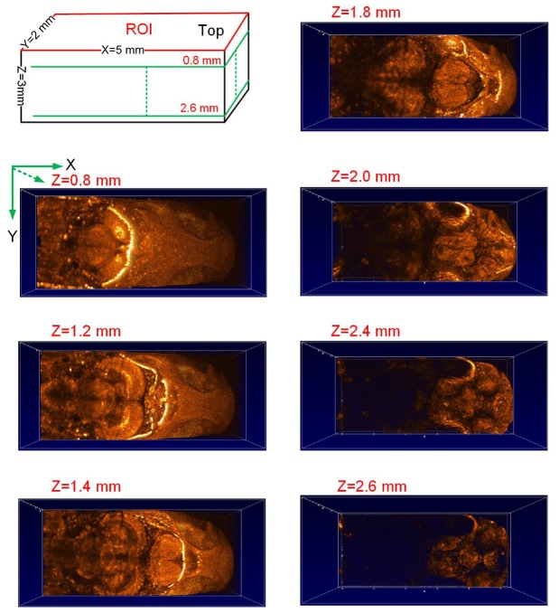
Fig. 6.
Characterization of the adult zebrafish brain from the horizontal view based on the reconstructed 3D SD-OCT image of the same region of interest (ROI) as shown in Fig. 4 (a) with different imaging depths along Z axis with z = 0.8 mm, 1.2 mm, 1.4 mm, 1.8 mm, 2.0 mm, 2.4 mm and 2.6 mm, respectively.

