
Fig. 3.
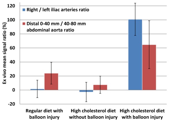
Signal comparison for selected arterial segments in ex vivo near-infrared fluorescence (NIRF) images. The distal 0-40 mm abdominal aorta and the right iliac artery are the balloon-injured regions, when performed. The ratios were obtained by dividing the mean right iliac artery (or distal 0-40 mm abdominal aorta) signal by the mean left iliac artery (or distal 40-80 mm abdominal aorta) signal for each animal. The error bars are the standard errors on the mean for the three rabbits of each group.