
Fig. 7.
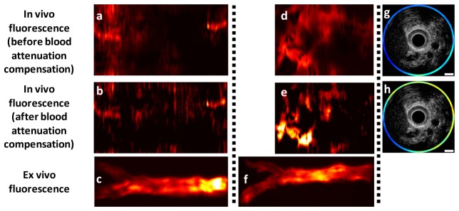
Effect of blood attenuation compensation on in vivo fluorescence images. (a, d, g) In vivo fluorescence images of the abdominal aortas of 3 rabbits without blood attenuation compensation; (b, e, h) Corresponding in vivo fluorescence images after blood attenuation compensation; (c, f) Corresponding ex vivo fluorescence images; (g, h) Integrated intravascular ultrasound (IVUS)/near-infrared fluorescence (NIRF) cross-sectional images (g) without and (h) with blood attenuation compensation.