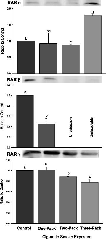
Fig. 2.

Western Blot Analysis for RAR Levels. Protein expressions of retinoic acid receptors (RARs: RARα, β, γ) in lungs exposed to different doses of cigarette smoke for 6 weeks. Representative Western Blot analyses are shown that used anti-RARα, anti-RARβ and anti-RARγ antibodies. The size of the detected RARα, RARβ and RARγ were all 53 kilodaltons (kDa). The intensity of the protein signal was determined by densitometry analysis (three samples in each group). The relative protein values in the three treatment groups were calculated as a mean value ± standard deviation (SD) of the data relative (ratio) to control with the control set at 1.0. Different letters represent significant difference among groups (P < 0.05)
