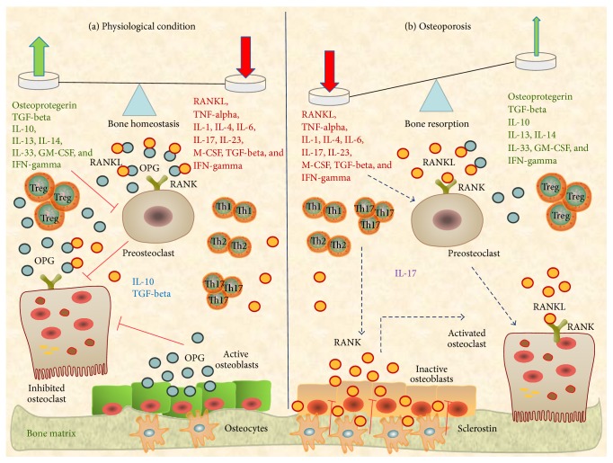
Figure 2.
Osteoclastogenic pathways in “bone immunological niche.” At bone tissue level, on one hand, there are the proinflammatory Th1, Th2, and Th17 cells with related proosteoclastogenic cytokines (RANKL, TNF-alpha, IL-1, IL-4, IL-6, IL-17, IL-23, M-CSF, TGF-beta, and IFN-gamma) and other immune cells such as proinflammatory macrophages; on the other hand, there are the anti-inflammatory Tregs with related antiosteoclastogenic cytokines (OPG, TGF-beta, IL-10, IL-13, IL-14, IL-33, GM-CSF, and IFN-gamma) and other immune cells with regulatory functions, such as osteomacs. The bone microenvironment functions as a dynamic model in which a continuous balancing between proosteoclastogenic and antiosteoclastogenic mediators is performed. (a) In physiological condition anti- and proosteoclastogenic factors are in equilibrium and bone homeostasis is conserved. (b) In osteoporosis condition, proosteoclastogenic factors prevail and bone resorption and remodeling develop. OPG: osteoprotegerin, GM-CSF: granulocyte-macrophage colony stimulating factor, and M-CSF: macrophage colony stimulating factor.

