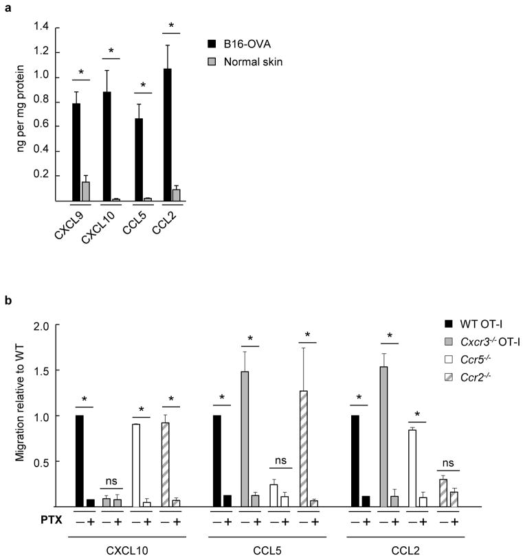
Figure 1. Murine CD8+ effector T cells express an array of functional chemokine receptors complementary for chemokine ligands present in the tumor microenvironment.
(a) Cognate chemokine ligands for CXCR3, CCR5, and CCR2 (i.e., CXCL9/CXCL10, CCL5, and CCL2, respectively) were quantified by ELISA in B16-OVA tumor extracts (tumor volume ~ 400–500 mm3) or in normal skin fom tumor-free mice. Data (mean ± s.e.m.) are from ≥3 independent experiments (n ≥2 mice per group). (b) Transwell assays were performed where fluorescently-labeled WT OT-I T cells were admixed with equivalent numbers of chemokine receptor-deficient effector cells and tested for migration to the indicated recombinant chemokines. WT and chemokine receptor-deficient cells were also pretreated with the global G-protein inhibitor pertussis toxin (PTX) and migration was quantified by flow cytometry. Background migration (absence of chemokine) was subtracted from all values. Data (mean ± s.e.m.) are represented as migration relative to WT and are from ≥3 independent experiments. (a, b) * P < 0.05; ns, not significant; unpaired two-tailed Student’s t-test.

