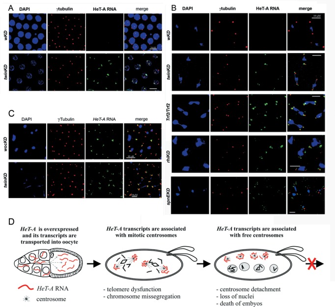
Figure 5.

Telomeric transcripts are localized around centrosome in syncytial embryos. Confocal images of 0−2-h-old embryos with the indicated genotypes. RNA FISH of HeT-A antisense probe revealed HeT-A RNA (green) near centrosomes in the prophase of twinKDnos embryos (A), during metaphase-anaphase in twinKDnos, Trf2/Trf2, rhiKDnos and spnEKDnos embryos (B) and in the cortex of wocKDnos and twinKDnos embryos (C). Red: γ-tubulin; blue: DNA. Bar: 10 μm. (D) The scheme depicts mitotic defects in early developmental stages as a result of HeT-A derepression in the germline and the accumulation of abundant HeT-A transcripts around centrosomes in embryos.
