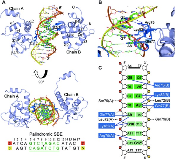
Figure 2.

Crystal structure of Smad5-MH1 in complex with the palindromic SBE sequence. (A) Overall structure of the Smad5-MH1/palindromic SBE complex. The Smad5 MH1 domains are colored in light blue, with the β-hairpin highlighted in marine. The zinc atoms are shown as spheres. The template chain and the complementary chain of the dsDNA are shown in orange and yellow, respectively. The DNA bases of the central SBE site are shown as sticks and colored in green. The DNA sequence used for crystallization is shown below the structure, with the central 8-bp palindromic SBE site underlined and highlighted in green. (B) Base-specific interactions by the β-hairpin. Residues and DNA bases involved in specific binding are colored as marine and green sticks, respectively. Hydrogen bonds are represented by blue dashed lines. (C) Diagram summarizing Smad5-MH1/SBE interactions. Non-interacting nucleotides are omitted. Each copy of the SBE motif is shaded in dark green and light green, respectively. The conserved β-hairpin residues, Arg75, Lys82 and Gln77, are shaded in blue. Interactions between the amino acids and DNA bases are shown as blue solid lines, while the contacts with the DNA phosphates are shown as black dashed lines.
