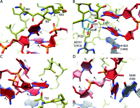
Figure 5.

Interaction of eRF1 with the UAA(A) stop codon. (A) The Uracil 1 (U1) nucleotide of the stop codon interacts with N7 of adenosine 3 (A3) and the A3 backbone phosphate to adopt U-turn geometry of the stop codon. Further, U1 interacts with N61 and K63 of the eRF1 TAS-NIKS motif. The hydroxylation site of K63 is located at C4 (*). (B) Adenosine 2 (A2) interacts with Hs A1825 (h44) and C127 (YxCxxxF motif). Possible rotamer conformations of E55 (E55 and E55#) for the interaction with A2 are depicted in light blue. Y125 (YxCxxxF motif) stabilizes the shifted conformation of Hs A3731 (H69). (C) A3 mainly interacts with T32 (GTS motif) and the 2′OH of U1. (D) Adenosine 4 (A4) stacks on the rRNA base Hs G626 (Ec G530). Interactions are indicated by dotted lines.
