
Figure 5.
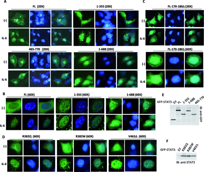
The intracellular localization of STAT3 truncations and mutants. (A and B) GFP-tagged STAT3-FL or indicated STAT3 domains were transfected into HepG2 cells. After transfection for 24 h, the cells were treated with or without IL-6 for 30 min and then fixed and visualized with a confocal microscope. Nuclei were stained with DAPI (blue). (C and D) GFP-tagged STAT3 mutants were transiently expressed in HepG2 cells. After transfection for 24 h, the cells were treated with or without IL-6 for 30 min and then fixed and visualized with a confocal microscope. Nuclear DNA was stained with DAPI (blue). (E) GFP-tagged STAT3-FL or indicated STAT3 domains were transfected in 293T cells. After transfection for 36 h, the cell lysates were detected by immunoblotting with anti-GFP antibody. (F) GFP-tagged STAT3-wild-type (WT) and mutants were transiently transfected into 293T cells. After 36 h, the cell lysates were detected by anti-STAT3 immunoblotting.