
Figure 3.
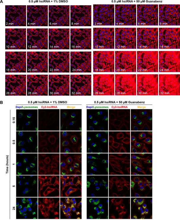
Guanabenz markedly impacts the rate and degree of intracellular uptake of hsiRNAs. (A) Uptake kinetics of hsiRNA. Cy3-PPIB hsiRNA (red), 0.5 μM, was added to HeLa cells in the presence of 1% DMSO or Guanabenz (50 μM). Imaged on a Leica confocal microscope, 63X, nuclei stained with Hoechst dye (blue). Laser intensity and acquisition time were fixed at the 2 min timepoint and kept constant throughout the experiment, resulting in saturation at later Guanabenz treatment timepoints. (B) Co-localization of hsiRNA with lysosomal markers. Cy3-HTT hsiRNA (red), 0.5 μM, was added in HeLa cells in presence of 1% DMSO or Guanabenz (50 μM). Nuclei stained with Hoechst dye (blue) and lysosomes stained with LysoTracker (green, Life Technologies). Cells were imaged on a Leica confocal microscope, with laser intensity and acquisition optimized for a 24 h experiment. Representative images taken at time points indicated are shown.