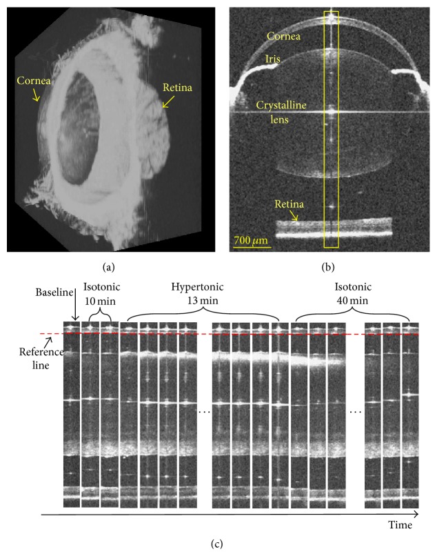
Figure 1.
OCT full-eye imaging and dynamic response to hypertonic challenge and reversal in mouse model. (a) Three-dimensional rendering of the OCT full-eye imaging of mouse. (b) Representative OCT cross-section image in the horizontal meridian. As marked by the yellow rectangular region in (b), a serial of clips around the corneal vertex reflection line were extracted and displayed along the time dimension to demonstrate the dynamic response in (c). In the depth direction, all clips were aligned along the posterior cornea.

