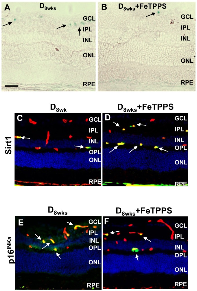
Fig 10. Senescence-associated retinal changes with FeTPPS treatment.
A,B) Retinal frozen sections from diabetic and diabetic + FeTPPS groups were probed at pH6 for detection of SA-β-gal positivity (blue, black arrows). Representative images from frozen retinal sections probed with anti-SIRT1 (C, D) and anti-p16INK4a (E, F) (green) to detect immunoreactivities in diabetic and retinas of FeTPPS- treated diabetic rats. Sections were co-labeled with anti-isolectin B4 (vascular structures, red). Hoescht staining was used to detect nuclei (blue). Scale bar equal to 50μm.

