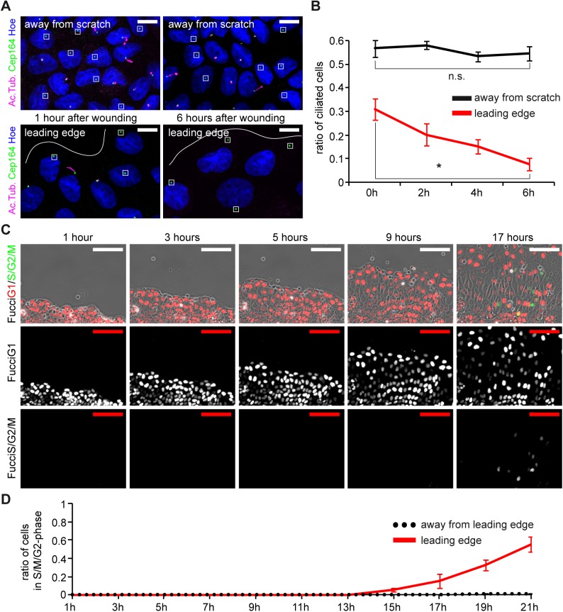
Fig 2. MDCK cells lose cilia during sheet migration.
(A) Confluent layers of ciliated MDCK cells (7 days after seeding) were scratched, fixed immediately (0h) or 6h after wounding, then stained with antibodies against acetylated Tubulin for cilia (magenta), Cep164 for the basal body (green) and Hoechst for nuclei (blue). Cilia occur at different lengths, basal bodies devoid of magenta are unciliated (squared). White lines indicate the leading edge. Scale bars: 10μm. (B) Quantification of ciliated cells at various time points in areas away from the leading edge and at the leading edge. 0h: 56.7%±3.3% vs. 31.0%±4.5%, 2h: 58.1%±1.7% vs. 20.2%±4.7%, 4h: 53.3%±2.1% vs. 15.0%±3.0%, 6h: 54.6%±3.1% vs. 7.5%±2.7% (asterisk: p < 0.01). n = 10 fields of view in two independent experiments. Note that the number of cilia at time point 0h at the leading edge is decreased compared to distant cells due to mechanical injury from wounding. (C) Analysis of proliferation in cells after wounding. MDCK cells stably expressing Fucci cell-cycle indicators were monitored after 7 days post seeding after scratch wounding. Nuclei of non-proliferating cells in G1 express RFP (red), nuclei of proliferating cells (S/G2/M) express GFP (green). Scale bars: 100μm. (D) Quantification of proliferating cells at the leading edge and cells away from the wound over 21 hours after scratch. n = 10 fields of view from two independent experiments.

