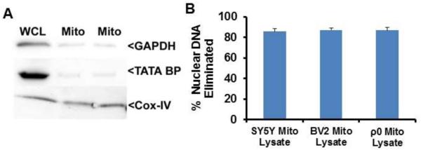
Fig. 1.

Characterization of mitochondrial lysates. (A) 50 μg of whole cell lysate (WCL) or mitochondrial lysate were resolved by electrophoresis and transferred to a nitrocellulose membrane. Representative immunochemistry data for COX-4I1 (mitochondrial marker), TATA BP (nuclear marker), and GAPDH (cytosolic marker) protein are shown. (B) The cycle threshold difference between DNA from whole cells and DNA from enriched mitochondria was used to calculate the amount of nuclear DNA that was eliminated during the mitochondrial enrichment process.
