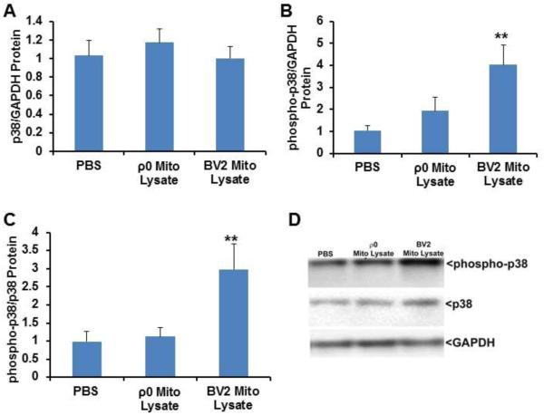
Fig. 4.
Effect of mitochondrial lysates on microglial cell p38 protein and phosphorylation levels. BV2 cells were treated overnight with PBS vehicle, 50 μg of mitochondrial lysate from BV2 cells, or 50 μg of mitochondrial lysate from SH-SY5Y ρ0 cells. Cells were lysed using MPER, equal protein concentrations were resolved by electrophoresis, gel proteins were transferred to nitrocellulose membranes, and the membranes were immunoblotted for p38, GAPDH, and phosphorylated p38 protein. (A) Total p38 normalized to GAPDH. (B) Phosphorylated p38 normalized to GAPDH. (C) Phosphorylated p38 normalized to total p38. (D) Representative immunochemistry data. Values shown are relative group means ± SEM, with the control group set to 1. ** indicates p<0.005.

