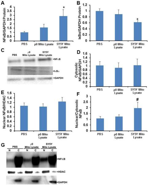
Fig. 6.
Effect of mitochondrial lysates on neuronal cell NFκB protein levels and compartmentalization. SH-SY5Y cells were treated overnight with PBS vehicle, 50 μg of mitochondrial lysate from SH-SY5Y cells, or 50 μg of mitochondrial lysate from SH-SY5Y ρ0 cells. Ten to twelve independent samples were prepared and analysed for each condition. (A) Total cell NFκB protein. (B) Total cell IκBα protein. (C) Representative whole cell immunochemistry data. (D) Cytosolic NFκB protein. (E) Nuclear NFκB protein. (F) Nucleus:cytosol NFκB protein ratio. (G) Representative nucleus and cytosol immunochemistry data. Values shown are relative group means ± SEM, with the control group set to 1. * indicates p<0.05; # indicates a non-significant ANOVA, but with p<0.05 between the control group and the indicated group on LSD post hoc testing.

