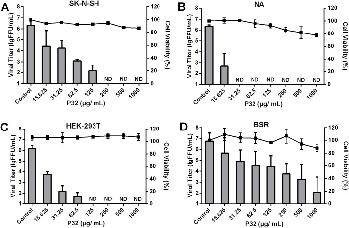
Fig 2. Cytotoxicity and RABV inhibitory effect of P32 on different cell lines.
To assess the cytotoxicity of P32 in different cell lines, we used the MTS method. SK-N-SH (A), NA (B), HEK-293T (C), and BSR cells (D) were incubated with different concentration of P32 for 48 h at 37°C, after which the cells were incubated with MTS solution for 1 h. Absorbance at 490 nm was determined, and the percent cell viability for each P32 treatment group was determined by comparing the treated cells with mock-treated cells. For the inhibition assay, cells were infected with SAD-L16-eGFP at an MOI of 0.01 after a 2-h incubation with P32. Supernatants were harvested for virus titration at 48 hpi. Columns and black solid squares represent the viral titers and the cell viability percentages, respectively. Each value is expressed as the mean ± SEM from three independent experiments. ND means non-detectable.

