
Fig 6. P32 affects cell fusion mediated by RABV G protein.
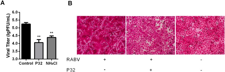
(A) Viral titers in response to P32 treatment during the post-internalization period. NA cells were infected with SAD-L16 at an MOI of 0.01 and subjected to the post-internalization inhibition assay, after which viral titers were determined. Each value is expressed as the mean ± SEM from three independent experiments (*, P<0.05; **, P<0.01; ***, P<0.001; ****, P<0.0005). (B) P32 inhibits G protein-mediated cell membrane fusion. The low pH-dependent membrane fusion assay was performed in RABV-infected BSR cells. Briefly, BSR cells were infected with SAD-L16 at an MOI of 5 at 37°C for 48 h. After viral infection, cells were treated with MM containing P32 or vehicle at 37°C for 2 h, after which the cells were washed and incubated with fusion medium for 2 min at room temperature. Then, the fusion medium was discarded, and MM was added to the cells, which were incubated at 37°C for another 2 h. The cells were then fixed with 80% ice-cold acetone and stained with fuchsine solution.