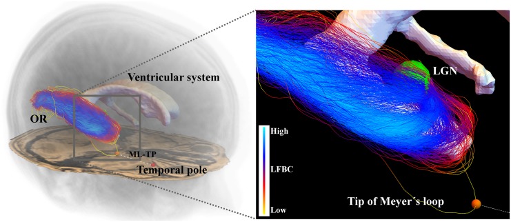
Fig 12. A reconstruction of the optic radiation and its positioning in the brain.
The left figure shows how the OR is positioned in the brain, the close-up on the right shows how the OR wraps around the ventricular system. The probabilistic tractography outputs many spurious fibers. The tip of the Meyer’s loop, indicated by the orange sphere, is localized on a spurious fiber and is therefore very dependent on the realization of the tractography. As a result, the distance from the Meyer’s loop to the Temporal pole (ML-TP) that is used in temporal lobe resection surgery, shows a high variation among different tractography outcomes.

