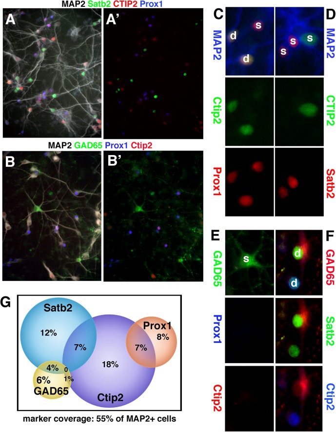
Fig 3. Non-exclusivity in expression of transcription factors used as cell type markers.
(A,B) Quadruple stainings of DIV9 rat hippocampal neurons with MAP2 (white), Ctip2 (red), Satb2 (green), and Prox1 (blue) (A), or MAP2 (white), GAD65 (green), Prox1 (blue), Ctip2 (red) (B). (A’) and (B’) show the same field with MAP2 staining omitted. (C-F) show examples of single (s) and dually (d) positive neurons. (C) Ctip2/Prox1-single (s) and dually (d) positive neurons. (D) single-positive Ctip2- or Satb2-positive neurons. (E) Gad65-positive neuron not expressing Prox1 or Ctip2. (F) GAD65/Satb2 double-positive neuron and Ctip2/Satb2 double-positive neuron. (G) Venn diagram showing the abundance of single and double-positive neurons. Two independent DIV9 hippocampal cultures were scored and normalized to MAP2-positive cells. In each experiment, 300–400 MAP2-positive neurons were counted.

