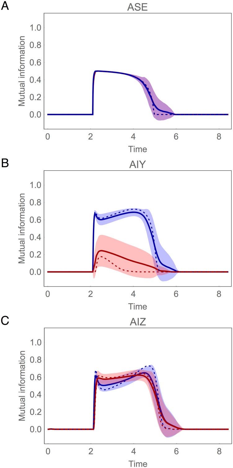
Fig 12. Mutual information during concentration step assay in the population of successful circuits.
(A) Chemosensory neuron ASE: left (blue) and right (red) cell. (B) Interneuron AIY: cell with highest mutual information in blue, the other cell in red. (C) Interneuron AIZ: cell downstream from the AIY cell with highest mutual information in blue, the other cell in red. Mean (solid trace) and standard deviation (shaded area) for the ensemble of successful networks. Mutual information for the best circuit shown as a dotted trace.

