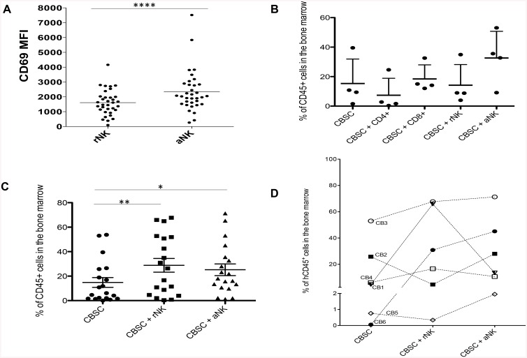
Fig 1. Level of hCD45+ Cell Engraftment in the BM and Fold Increase in Engraftment Observed in NSG Mice Transplanted with CBSC and CB NK Cells.
(A) CD69 expression measured as mean fluorescence intensity (MFI) on CB NK cells before and after incubation with 20 ng/mL IL-15 for 4 h (N = 18). (B) Percentage of hCD45+ cells detected by flow cytometry in the BM of NSG mice ten weeks post-transplant with CBSC alone or in combination with CD4+ T cells, CD8+ T cells, rNK cells or aNK cells (N = 4). (C) Percentage of hCD45+ cells detected by flow cytometry in the BM of NSG mice ten weeks post-transplant with CBSC alone or in combination with rNK cells or aNK cells (N = 14). (D) Percentage of hCD45+ cells detected by flow cytometry in the BM of NSG mice transplanted with CBSC alone or in combination with rNK cells or aNK cells from 6 different CB units (N = 18). * P < 0.05, ** P < 0.01, *** P < 0.001.

