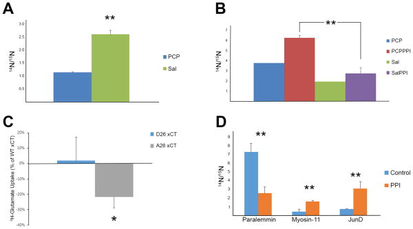
Figure 4.
A) The peptide ADSpFSEGDDLSQGHLAEPCFLR of Begain was significantly down-regulated after PCP treatment. B) The peptide LPpSVGDQEPPGHEK of xCT was significantly up-regulated after PCPPPI. The same trend was trend observed for the PCP and Sal comparison but not enough measurements were collected for statistical analysis. C) Abolishing the phosphorylation site in B) decreases 3H-glutamate uptake through xCT in HT22 cells. Cells were transfected with WT, D26, or A26 xCT cDNA and glutamate uptake assays was performed. The data represents three independent experiments. The y-axis is the 3H-glutamate measurements of D26 and A26 as a percentage of the WT xCT glutamate uptake measurements. D) Phosphopeptides significantly altered in the PFC between rats that underwent PPI or mock PPI (control). Paralemmin (SETMVNAQQpTPLGpTPK), myosin-11(VIENTDGpSEEEMDAR), JunD (LAALKDEPQTVPDVPSFGDpSPPLpSPIDMDTQER). * p value < 0.05, ** p value< 0.01.

