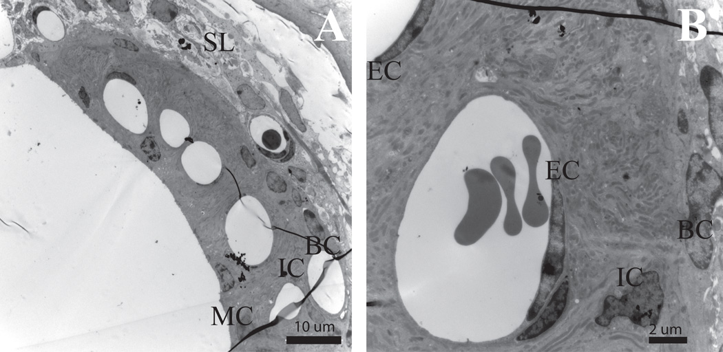
Figure 3. Stria vascularis (SV) shows no radiation-induced changes in the 60 Gy group.
(A) Tissue of the cochlear lateral wall shows no evidence of edema, vacuoles or nuclear condensation in either the fibrocytes of the spiral ligament or the three cell layers of the stria vascularis. Normal capillary density is evident in this mid-modiolar cross-section of the 60 Gy SV at 8 days post-irradiation. (B) Normal capillary wall endothelium and surrounding SV in a control mouse. (C) Normal capillary wall endothelium and surrounding SV in 60 Gy mouse at 8 days post-irradiation. SL = Spiral Ligament, BC =Basal Cell, IC =Intermediate Cell, MC =Marginal Cell, EC = Endothelial Cell, PC= Pericyte, Cap=Capillary Scale bar A=10 µm, B=2 µm

