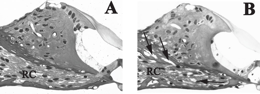
Figure 4. Axonal density traversing spiral lamina is decreased in the 60 Gy group.
(A) The auditory nerve fibers in the spiral lamina of a control mouse show minimal area unoccupied by bipolar axons or extracellular matrix. (B) The spiral lamina of a mouse 8 days post-irradiation with 60 Gy has areas devoid of axons, glia or extracellular matrix (arrows) at 8 days post-irradiation. Although the cells of the organ of Corti from spiral limbus to inner pillar cell appear normal, a vacuole (arrowhead) can be detected at the base of the inner hair cell. AN= auditory nerve fibers, SpL= spiral limbus, tm= tectorial membrane. Scale bar= 50 µm

