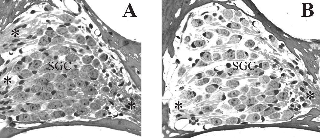
Figure 5. Spiral ganglion (SG) cell density is decreased in the 60 Gy group.
(A) Rosenthal’s canal in a control mouse shows a high density of spiral ganglion cells and associated axon bundles. (B) Decreased SG and extracellular matrix density is evident in the 60 Gy group at 8 days post-irradiation (arrows). Vacuoles are present in some of the remaining SG/satellite cell complexes (arrowheads). (C) An early marker of irradiation-induced cellular stress is vacuolization (arrowheads) in the satellite cells of a mouse at 8 days post-irradiation with 20 Gy. (D). Satellite cells (white dot on nucleus) show reduced cytoplasm and retraction (arrowheads) from their adjacent spiral ganglion cell. SGC=spiral ganglion cells, *= peripherally directed axons; **= centrally directed axons. Scale bar A&B= 20 µm, C&D = 5 µm

