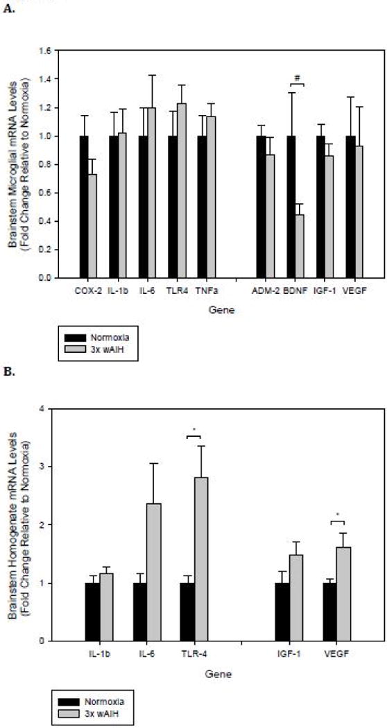
Figure 2. 3×W AIH does not promote inflammatory gene expression in rat brainstem microglia, but it increases expression of Tlr4 and Vegf in brainstem homogenates.

Gene expression was assessed by qRT-PCR in immunomagnetically-isolated microglia (A), and tissue homogenates from the brainstem (B) following 4 weeks of 3×W AIH. Treatment did not alter the expression of any inflammatory gene evaluated in microglia, though there was a non-significant reduction in Bdnf mRNA levels. 3×W AIH increased Tlr4 and Vegf gene expression in tissue homogenates. Data shown are means ± 1 SEM of n=5–8. *p<0.05 and #0.05<p<0.1 versus normoxia.
