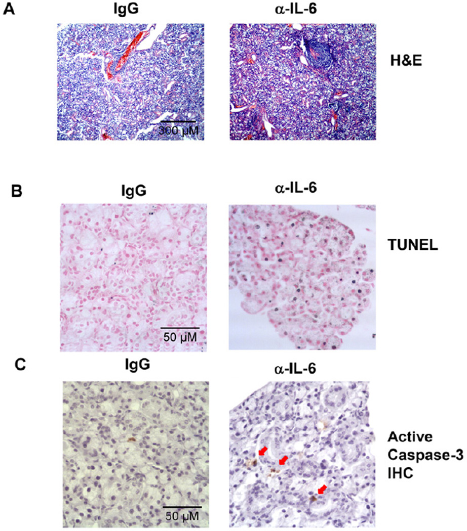
Figure 2. Neutralization of IL-6 in B6.NOD-Aec mice leads to increased inflammation and apoptosis in SMG.
Anti-IL-6 or control IgG was injected to 16 week-old B6.NOD-Aec mice, 3 times weekly for 8 weeks. (A) H&E staining of SMG sections showing leukocytic foci. Original magnification: ×10. (B) In situ TUNEL staining of apoptotic cells in SMG sections. Original magnification: ×40. (C) Immunohistochemical staining of active caspase-3 in SMG sections. Data are representative of the analyses of 9 mice for each group from 5 independent experiments. Original magnification: ×40.

