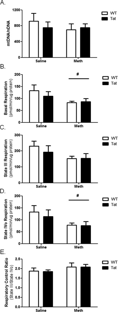
Figure 3. Cardiac mitochondrial dysfunction is caused by METH.
(A) Cardiac LV mtDNA abundance was determined for each experimental group and normalized to nuclear DNA. No change in LV mtDNA abundance was observed in any experimental group compared to LV of WT-saline mice. Mitochondria were isolated from LV tissue and analyzed using a Seahorse XF24 oximetric analyzer. Basal respiration (B) was significantly reduced in METH-treated mice. State III respiration (C) was not significantly altered following METH exposure, but State IVo respiration (D) was significantly reduced following METH exposure. The respiratory control ratio (E) was unchanged. Tat transgene did not significantly alter any mitochondrial functional parameter within the experimental timeline. # denotes p<0.05 for METH-treated groups compared to saline-treated groups by two-way ANOVA.

