. 2015 May 29;7(6):557–564. doi: 10.4168/aair.2015.7.6.557

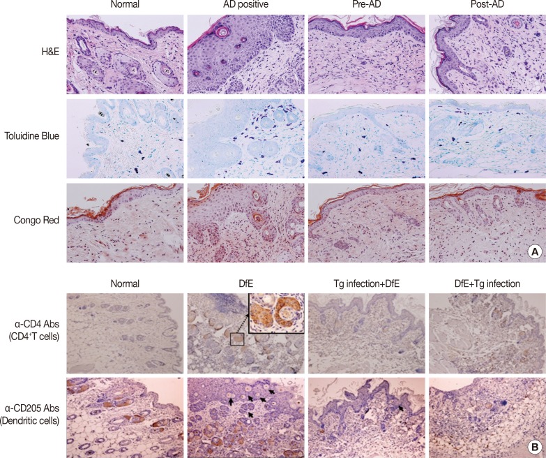
Fig. 2. T. gondii suppressed infiltration of immune cells in the skin of mice with atopic dermatitis. (A) Hematoxylin-eosin staining of the lesioned skin revealed heavy inflammation, hyperkeratosis, acanthosis, and parakeratosis. Infiltration of inflammatory cells into the skin after DfE application was determined by staining tissue sections with toluidine blue or Congo red to detect mast cells and eosinphils, respectively. Original magnification,×200. (B) Immunohistological staining revealed infiltration of CD4+ T cells and dendritic cells in the dermis/epidermis. Paraffin sections were immunostained with anti-CD4 or anti-CD205 antibody to detect CD4-positive cells and dendritic cells, respectively. Positively stained cells are shown in brown color. Arrows indicate dendritic cells in the epidermis. Original magnification,×100. Results shown represent three independent experiment (n=5 for each group). Tg, T. gondii; DfE, Dermatophagoides farinae extract.

Fig. 2