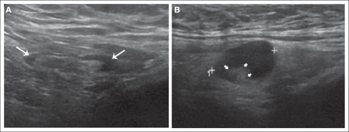
Fig 1.
(A) Illustration of normal lymph nodes on ultrasound. Ultrasound image of a morphologically normal lymph node with uniform thin hypoechoic cortex (white arrows) less than 3 mm in thickness. (B) Illustration of abnormal lymph nodes on ultrasound. Ultrasound image of a metastatic axillary lymph node with diffuse hypoechoic cortical thickness and deformity of the echogenic fatty hilum (small white arrowheads).

