
FIGURE 2.
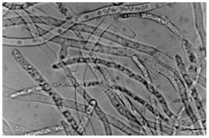
Microscopic (40×) observation of Sclerotium rolfsii ATCC 201126 mycelium after growth for 3 days at 30°C in liquid PM20 (with 20 g/L sucrose, Fariña et al., 1998). Mycelium was disaggregated in 10% w/v KOH.
Official websites use .gov
A
.gov website belongs to an official
government organization in the United States.
Secure .gov websites use HTTPS
A lock (
) or https:// means you've safely
connected to the .gov website. Share sensitive
information only on official, secure websites.
Microscopic (40×) observation of Sclerotium rolfsii ATCC 201126 mycelium after growth for 3 days at 30°C in liquid PM20 (with 20 g/L sucrose, Fariña et al., 1998). Mycelium was disaggregated in 10% w/v KOH.