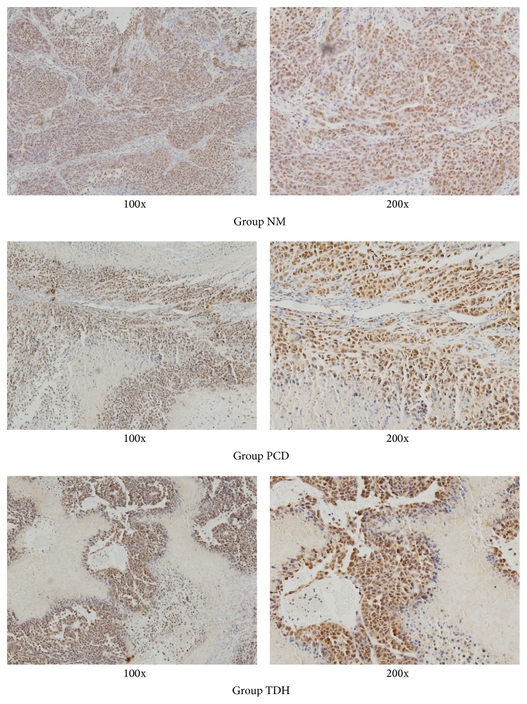
Figure 6.
PCNA expression in tumor tissues assayed with immunohistochemical staining. Immunohistochemistry was performed to measure PCNA in tumor tissues derived from control and treated mice (magnification 100x and 200x). 5 FU and TDH were dissolved by saline to certain concentration. The dosage of drugs was as follows: in Group NM, saline 20 mL·kg−1; in Group PCD, 5 FU 20 mg·kg−1; in Group TDH, TDH 100 mg·kg−1. Cells characterized with positive expression of PCNA took on brown. Cells with negative took on blue.

