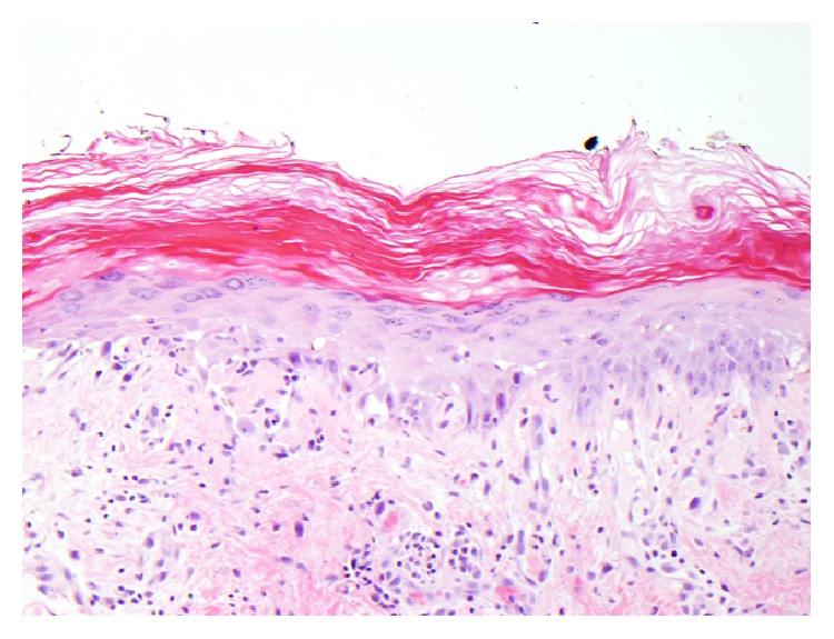
Figure 3.

Figure showing skin biopsy of the lesion. Vacuolar alteration of the basal layer and numerous clumped cytoid bodies along the dermoepidermal junction and focally within the spinous layer, stratum corneum, and superficial adnexal epithelium are seen. Within the dermis, there is a superficial and mid perivascular and focally perifollicular inflammatory infiltrate comprised predominantly of lymphocytes with scattered melanophages. Neutrophils and eosinophils are not conspicuous.
