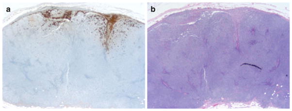
FIG. 1.

a S100 immunohistochemistry stain (×25): positive for metastatic melanoma at periphery of lymph node. b H&E stain (×25): lymph node with effacement of nodal architecture and replacement by diffuse growth of small, monotonous lymphocytes, as well as microscopic involvement by metastatic melanoma
