Fig. 6.

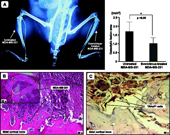
Everolimus restrains the bone-metastatic potential of BC cells in vivo. Everolimus-treated and untreated MDA-MB-231 cells were intra-tibially inoculated in 8-week old SCID mice. a X-Ray image of a representative SCID mouse after 4 weeks, showing blown cortical bone of the right tibia and a smaller bone lesion produced by Everolimus-treated MDA-MB-231 cells in the left tibia. The graph shows levels of tibial erosion, measured by ImageJ software. Data are expressed as mean ± SE of the 2D size of metastatic lesions (untreated MDA-MB-231: 1.7 ± 0.56 mm2; Everolimus-treated MDA-MB-231: 0.9 ± 0.31 mm2; *p < 0.05). b Representative image of tumor infiltration in an excised tibia (H/E staining). The small box includes a horizontal section of the right tibia, showing that the cortical bone is largely infiltrated by MDA-MB-231 cells, as revealed at higher magnitude. c Detection of TRAcP+ cells at the tumor/bone interface of the same tibia
