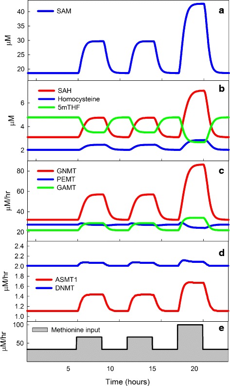
Fig. 7.

The effect of meals on competing methyltransferases. The input of methionine into the liver Panel e was varied during a 24 hour period: 33.33 μM/hr until breakfast, then 66.67 μM/hr for 3 hours, similarly for lunch, and for 3 hours after dinner the input is 100 μM/hr. Panel a shows the large deviations in SAM due to the methionine input changes. Panel b shows that SAH and Hcy track the changes in SAM, but 5mTHF has the opposite changes because SAM inhibits MTHFR (see Fig. 1). The changes in Hcy are small because SAM stimulates CBS. Panel c shows the time course of the fluxes through GNMT, PEMT, and GAMT. As explained in the text, GNMT goes up rapidly as SAM increases taking most of the extra methyl groups so that the changes to GAMT and PEMT are modest. The changes to PEMT are exceptionally small (see the text for Section The effect of inhibition by SAH). Panel d shows the fluxes through AS3MT and DNMT. The changes to DNMT are exceptionally small (see the text for Section The importance of small Km values) because the K m of DNMT for SAM is very small. By contrast, AS3MT has a much higher K m and therefore varies much more
