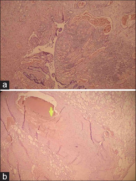
Figure 1.

(a) Photomicrograph of small intestine showing focal ulceration of the mucosa with marked inflammatory cell infiltration and vascular congestion (H and E, ×40). (b) Photomicrograph of mesentery showing thrombus (arrow) in the mesenteric artery (H and E, ×40)
