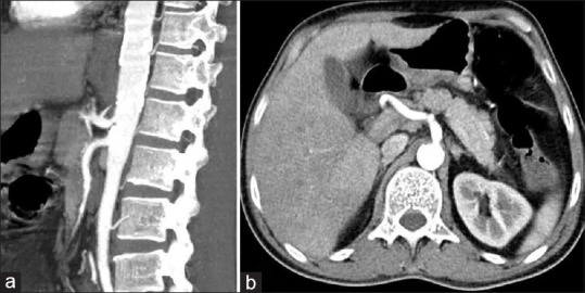
Figure 3.

Photographs of computed tomography abdomen angiography sagittal (a) and axial (b): Calcified plaques in the abdominal aorta with focal narrowing of the celiac trunk at origin approximately 50% seen. There is normal caliber and flow in proximal superior mesenteric artery (SMA) with irregular slight thickening of SMA wall causing narrowing of SMA approximately 60%, mesenteric fat stranding in lower abdomen
