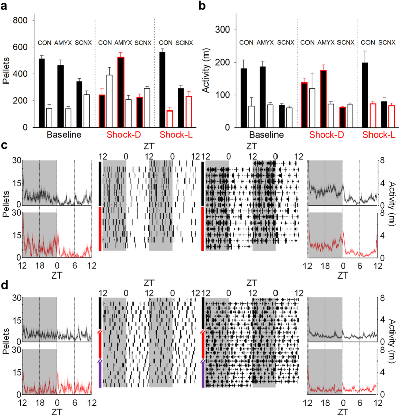
Figure 8. Effects of lesions on shock-induced changes to circadian rhythms.
(a) Mean number of pellets and (b) mean activity (distanced travelled in m) in the dark (filled bars) and light (open bars) phases of AMYX (n = 7), SCNX (n = 8), and sham (CON; n = 7) animals during the last 5 d of baseline (left), unsignaled nocturnal (center) and unsignaled diurnal shock (right; SCNX and CON only). Red highlights indicate shock period. Error bars represent SEM. (c) 24-h waveforms (outside) and raster plots (inside) of feeding (left) and activity (right) from amygdala-lesioned rats (n = 7; baseline and unsignaled shock conditions represented by black and red, respectively). (d) 24-h waveforms (outside) and raster plots (inside) of feeding (left) and activity (right) from SCN-lesioned animals (n = 8). Waveforms show the mean feeding and activity over 24 h (bold lines), in 10-min time-bins, of all animals in each group averaged over the last 5 d of each experimental condition. SEM is shown as the shaded areas above and below the bold lines.

