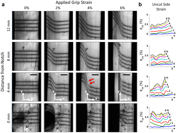
Figure 2. Notch tension testing of tendon fascicles.
(a) With increasing grip-to-grip strain, the notch widened and the photobleached lines became steeply angled, representing shear strains developing within the tissue. Consistently at 4% grip-to-grip strain, discontinuities in the photobleached lines first appeared at the location 4 mm from notch (red arrows). These discontinuities propagated parallel to the fascicle axis demarcating the interface between the cut and uncut portions of the tissue. (b) At all non-zero distances from the notch, the axial strains (εyy) on the uncut side of the tissue exhibit gradients similar to those seen in the gel (Fig. 1c). Scale bars, 200 μm.

