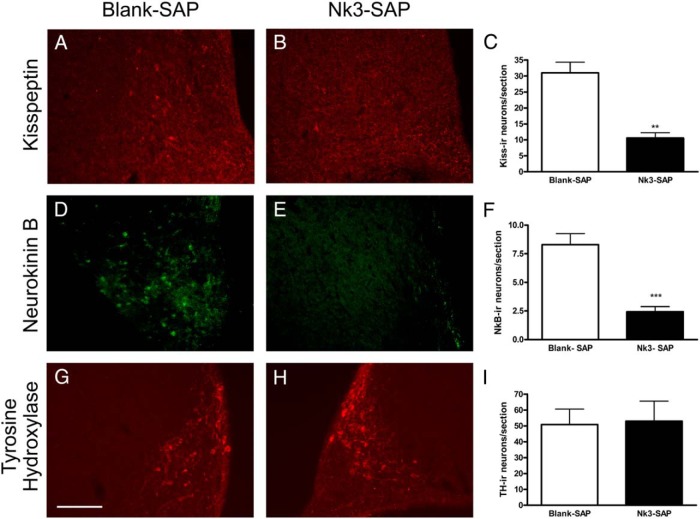
Figure 2.
Photomicrographs of coronal brain sections showing kisspeptin, neurokinin B, and tyrosine hydroxylase immunoreactivity in the arcuate region of rats submitted to microinjection of blank-SAP (A, D, and G) or Nk3-SAP (B, E, and H) after 7–10 days. Quantification of the number of kisspeptin (C), neurokinin B (F), and tyrosine hydroxylase (I) cell bodies in the arcuate region of the same animals. Data are shown as mean ± SEM. **, P < .01, ***, P < .001 compared with control. Scale bar 100 μm.

