Figure 3.
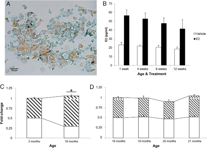
Vaginal cytology, E2 plasma concentrations, and alternative splicing of ERβ in intact animals. A, Representative image (×20) from vaginal smears that were obtained from 18-month-old female rats daily for 7 days before OVX (n = 6). Cells were stained with Papanicolaou stain before imaging. Orange G (orange) stains keratinized squamous epithelial cells. Eosin azure (blue) staining cells represent nonkeratinized squamous epithelial cells, neutrophils, and red blood cells (if present). B, Concentration (pg/mL) of plasma E2 in vehicle- and E2-treated animals during hormone-deprivation paradigm. E2 concentration was analyzed by E2 high-sensitivity ELISA kit; 18-month animals before OVX had low circulating E2 levels (35.0 ± 7.1 pg/mL; n = 6). C, Total ERβ (white region) and ERβ2 (hatched region) mRNA expression was measured in the hypothalamus of intact (non-OVX) 3 and (D) 18- to 21-month-old female rats. a, statistically significant difference (P < .05) in total ERBETA as determined by paired t test. Data are expressed as mean ± SEM.

