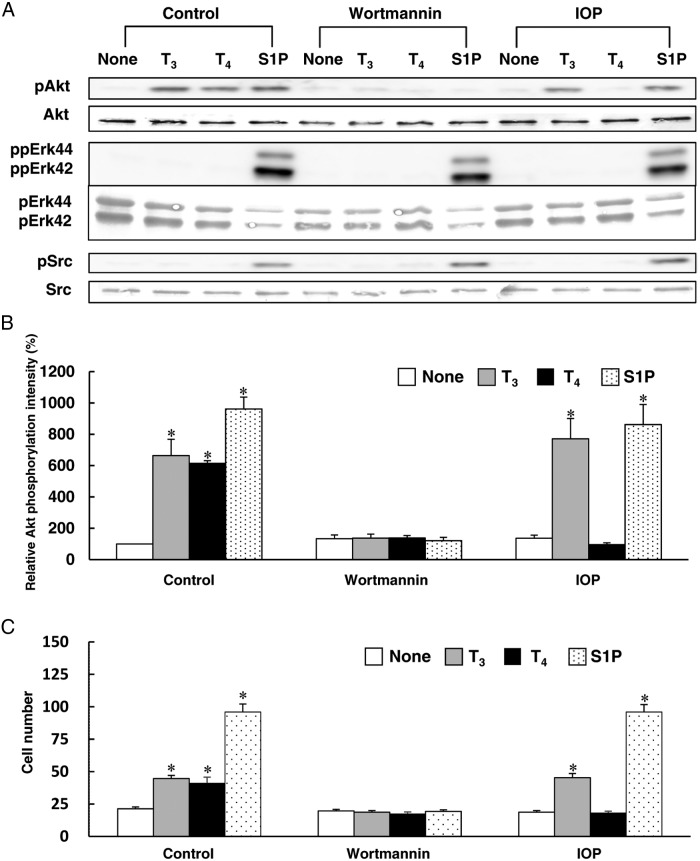
Figure 3.
The effects of wortmannin and IOP on TH-induced Akt phosphorylation and migration in HUVECs. Before performing the experiment described below, HUVECs were treated for 4 h at 37°C with RPMI 1640 medium containing 0.1% fatty acid–free BSA. A, HUVECs were pretreated with or without 100nM wortmannin or 1μM IOP for 20 min at 37°C in RPMI 1640 medium containing 0.1% fatty acid–free BSA. The cells were then stimulated for 10 min at 37°C with T3 (1nM), T4 (1nM), S1P (1μM), PBS (none), wortmannin, or IOP to measure the phosphorylated form and total Akt, pErk42/44 and Src by Western blotting. B, The relative amount of Akt phosphorylation was calculated from three independent experiments shown in panel A. C, HUVECs were pretreated with or without 100nM wortmannin or 1μM IOP for 20 min at 37°C in RPMI 1640 medium containing 0.1% fatty acid–free BSA. The cells were then stimulated for 4 h at 37°C with T3 (1nM), T4 (1nM), S1P (1μM), PBS (none), wortmannin, or IOP in RPMI 1640 medium containing 0.1% fatty acid–free BSA to investigate the migration of HUVECs using a blind Boyden chamber apparatus. The number of migrated cells attached to the lower surface of the filters was counted under a microscope at 400× magnification. Representative results of three separate experiments are shown in panel A. Results are presented as means ± SEM of three separate experiments in panels B and C. *, P < .05 vs none.

