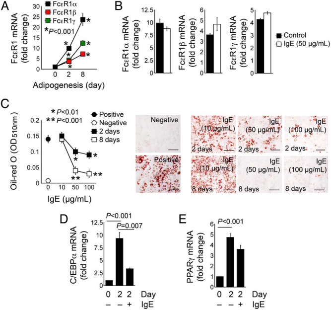
Figure 5.
IgE activity in reducing 3T3-L1 cell adipogenesis. A, RT-PCR determined the mRNA levels of three FcϵR1 chains (α, β, and γ) in 3T3-L1 cells at three time points (0, 2, and 8 d) during the differentiation. B, RT-PCR determined the expression of FcϵR1 three chains (α, β, and γ) in 3T3-L1 cells differentiated for 2 days with and without 50 μg/mL IgE. C, Oil-red O staining and quantification of 3T3-L1 cells treated with different doses of IgE for the first 2 days or throughout the whole course of differentiation (8 d). Preadipocytes and fully differentiated 3T3-L1 cells without IgE treatment were used as negative and positive controls. Representative data are shown to the right. RT-PCR determined the mRNA levels of C/EBPα (D) and PPARγ (E) in 3T3-L1 cells before differentiation and after 2 days of differentiation meanwhile treated with and without 50 μg/mL IgE. Data are mean ± SEM of three to five independent experiments.

