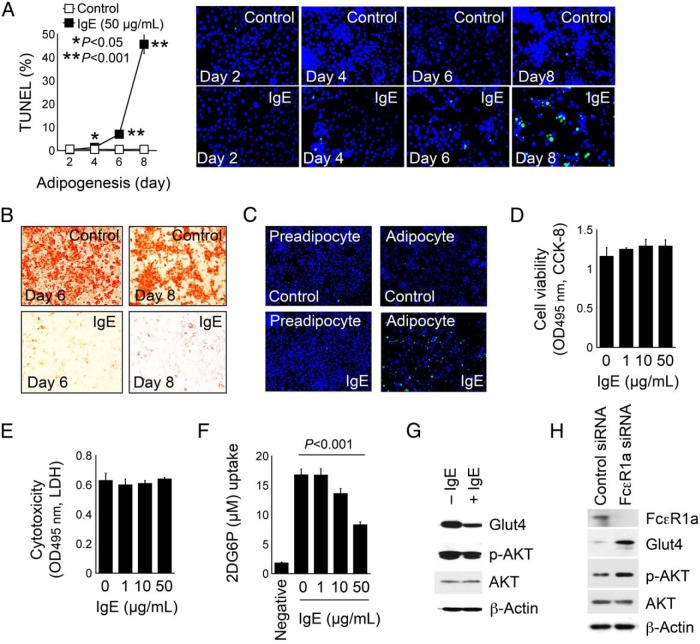
Figure 6.
IgE activities in promoting adipocyte apoptosis and suppressing adipocyte glucose uptake. A, TUNEL staining and quantification of apoptotic cells in 3T3-L1 cells differentiated in the presence and absence of 50 μg/mL IgE for indicated days. Representative data are shown to the right. B, Oil-red O staining of the same experiment from day 6 and day 8 experiments of panel A. C, TUNEL staining of preadipocytes and fully differentiated adipocyte treated with and without IgE (50 μg/mL) to induce cell apoptosis. A CCK-8 assay determined cell viability (D) and an LDH assay determined cytotoxicity (E) of 3T3-L1 cells after 2 days of differentiation with and without different amount of IgE. F, Glucose uptake assay of 3T3-L1 cells after 2 days of differentiation with and without different amount of IgE. Preadipocytes were used as an experimental negative control. G, Glut4, p-AKT, and total AKT immunoblots of differentiated 3T3-L1 cells and treated with and without 50 μg/mL IgE for 30 minutes. H, FcϵR1α siRNA- and scramble siRNA-transfected 3T3-L1 cells and differentiated for 4 days, followed by treatment with 25 μg/mL of insulin or IgE for 10 minutes. β-Actin immunoblots were used for protein loading controls. Data are mean ± SEM of three independent experiments.

