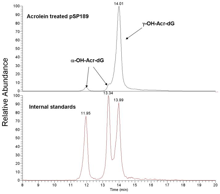
Figure 2.
Typical chromatograms obtained from LC-ESI-MS/MS analysis of Acr-modified pSP189 DNA. Double-stranded pSP189 DNA modified with different concentrations of Acr were denatured, digested with micrococcal nuclease, phosphodiesterase II, and alkaline phosphatase; the Acr-dG adducts were purified by SPE, and analyzed. Acr-dG adducts (upper trace) and standards of purified α-OH-Acr-dG and γ-OH-Acr-dG adducts (lower trace) was shown in. Note: two (+/−) stereoisomers of α-OH-Acr-dG were well separated while two (+/−) stereoisomers of γ-OH-Acr-dG cochromatographed at the same position (28).

