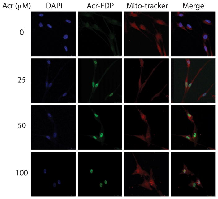
Figure 1.
Acrolein distribution in normal human lung fibroblasts (CCL202). Cultured CCL202 cells were treated with Acr (0–100 μM, 1 h), fixed, stained with anit-Acr-FDP-lysine (Nε-(3- formyl-3,4-dehydropiperidino)-lysine) antibody followed by goat anti-mouse FITC-conjugated secondary antibody, and then examined by microscopy. The method is the same as previously described [8]. Note: DAPI stained DNA, Acr-FDP stained Acr-lysine adducts and Mito-tracker stained mitochondria.

