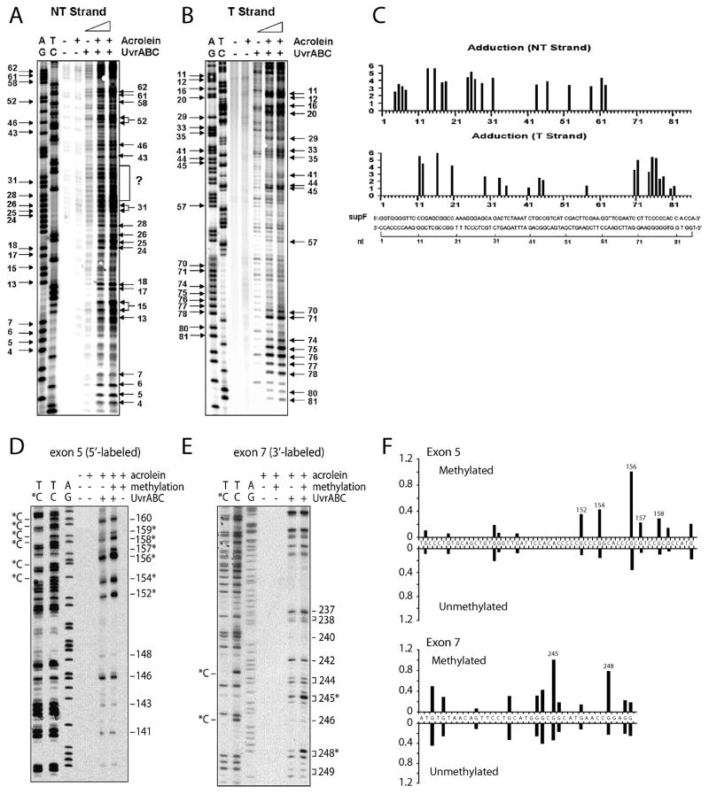
Figure 4.
Sequence specificity and cytosine methylation effect on Acr-dG adduct formation. A) and B) are 32P 5′-single end labeled supF DNA fragments. D) and E) are 32P 5′-single end labeled exon 5 of the p53 gene and 32P 3′-single end labeled exon 7 of the p53 gene fragments, respectively (with and without methylated at –CpG sequences by CpG methylase). Labeled DNA were modified with Acr and then reacted with UvrABC. The resultant DNAs were separated by denaturing DNA sequencing gel electrophoresis. C) and F) are quantitations. AG and TC represent Maxam and Gilbert sequencing reaction products; the positions of the methylated cytosines, which are resistant to Maxam Gilbert reactions, are indicated by C* [9, 11].

